
Persistent Neck Pain and Shoulder Asymmetry in a Child: A Diagnostic Challenge
Case Managed by – Dr. Ashish Naik, Dr. Yogin Patel and Dr. Pratik Bhujbal , Consultant Spine Surgeon, Alpha Ortho Spine Team
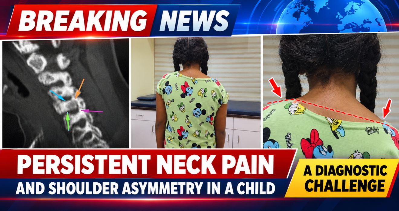
A 12-year-old girl presented with persistent neck pain, noticeable shoulder asymmetry and difficulty moving her neck. She had been experiencing these symptoms for several months and had already consulted multiple doctors, but the underlying cause had not been identified.
One detail stood out: her pain was particularly troublesome at night and improved with anti-inflammatory medication. These a symptoms called for a closer look at the cervical spine.
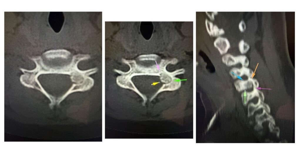
Advanced imaging eventually revealed a small bone lesion in the left C5 pedicle, part of the fifth cervical vertebra. The diagnosis was osteoid osteoma, a benign bone tumour that can cause persistent pain and, when it develops in the spine, may lead to muscle spasms, restricted movement and symptoms related to nearby nerves. This uncommon spine tumour in a child required careful assessment to determine the most appropriate treatment.
This rare spine case highlights why persistent neck pain in children requires careful assessment and how the right imaging and treatment strategy can help identify and manage an uncommon spinal condition.
When the Symptoms Don't Tell the Whole Story
Neck pain and shoulder asymmetry in a child can have several causes. Muscle strain and postural problems are among the possibilities, but persistent symptoms that do not settle deserve further investigation.
In this case, the combination of neck pain, restricted movement and pain radiating towards the left side made it important to look beyond the more common explanations.
The history of night pain was another significant clue. Osteoid osteoma is known for causing pain that often becomes worse at night and may respond well to NSAIDs. Recognising this pattern can help guide further investigations, particularly when symptoms have continued for months.
Why the CT Scan Was Important
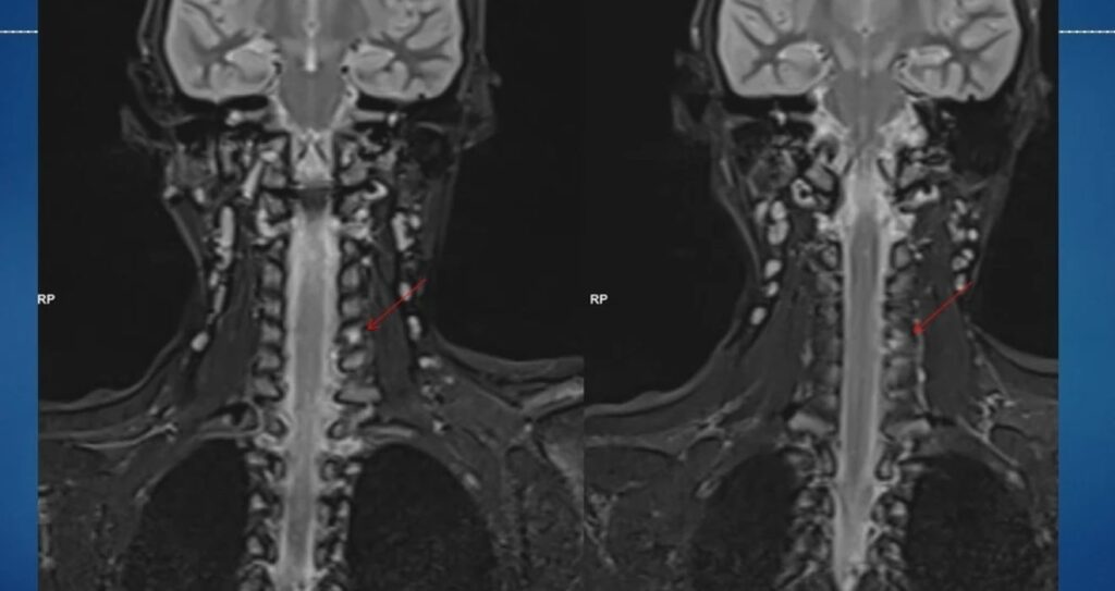
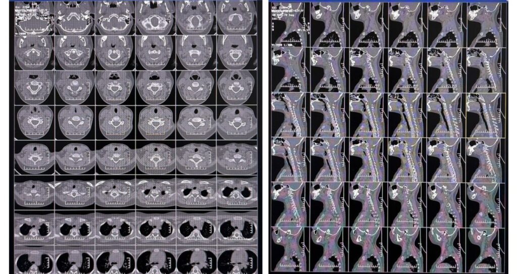
MRI is useful for assessing the spinal cord, nerve roots and surrounding soft tissues. However, osteoid osteoma can produce considerable inflammation in the surrounding bone. On MRI, this reactive oedema may be more prominent than the small lesion responsible for the symptoms.
CT imaging provides a detailed view of the bone and can help reveal the nidus, the small central area characteristic of osteoid osteoma.
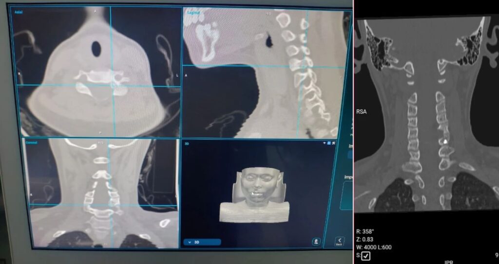
In this case, CT helped identify the lesion in the left C5 pedicle, providing an explanation for the patient’s persistent symptoms and supporting treatment planning. Accurate imaging is an important part of evaluating a suspected spinal tumour and planning appropriate spine surgery.
When a child’s symptoms do not correspond clearly with initial imaging findings, further assessment and appropriate imaging can help clinicians investigate uncommon causes of neck pain.
Choosing the Right Treatment for a Lesion Close to the Nerves
Once the diagnosis was established, the location of the lesion became an important consideration in deciding how to treat it.
Radiofrequency ablation (RFA) is a recognised treatment for osteoid osteoma. It uses heat to destroy the lesion without the need for conventional open surgery. However, the technique is not suitable for every case. When a lesion lies close to important neural structures, the risk of heat-related injury must be carefully assessed.
The C5 pedicle is located close to the spinal canal and adjacent nerve root. In this patient, that proximity influenced the decision to proceed with navigation-assisted spine surgery and surgical excision rather than RFA.
The aim was to locate and remove the lesion precisely while taking the surrounding anatomy into account.
The Alpha Ortho spine team selected this approach based on the lesion’s location and the need to consider the safety of nearby neural structures.
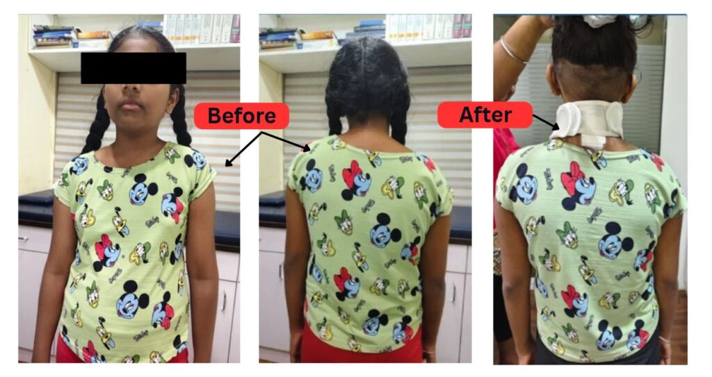
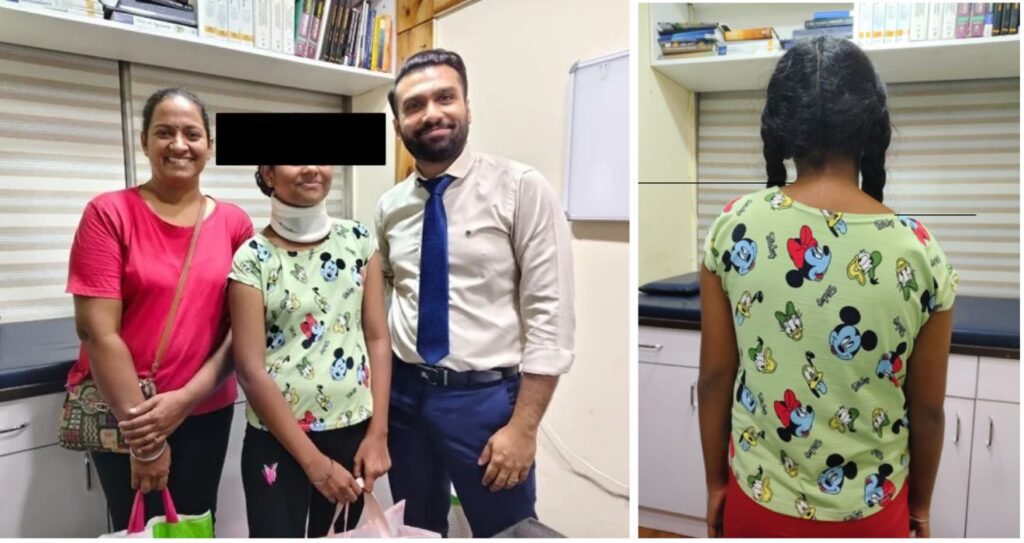
How Navigation-Assisted Surgery Helped
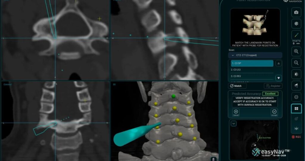
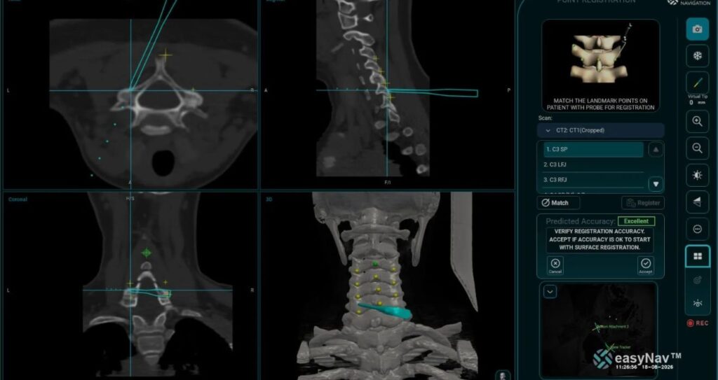
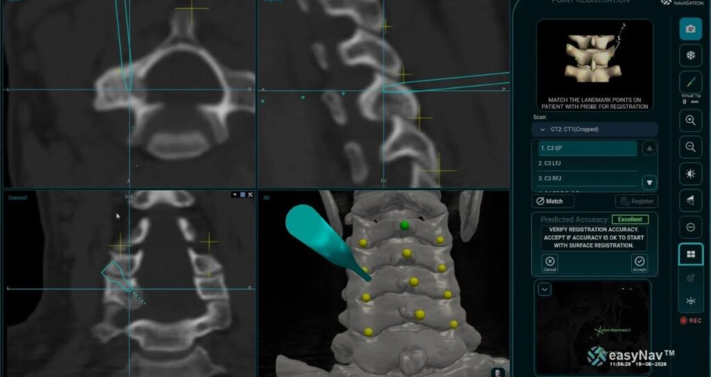
Navigation-assisted surgery uses imaging to help the surgeon identify the position of a lesion in relation to nearby structures. This can be particularly useful when operating in anatomically complex areas of the spine.
For this patient, navigation-assisted spine surgery allowed the lesion to be localised and removed with careful attention to the surrounding neural structures. The excised tissue could also be sent for histopathological examination to help confirm the diagnosis.
The procedure required a treatment plan tailored to the exact location of the lesion rather than a one-size-fits-all approach. Spine tumour surgery in children requires careful consideration of the lesion, the patient’s anatomy and the proximity of critical neural structures.
What This Case Teaches Us
The important lesson from this case is that persistent pain in a child deserves attention, especially when it follows an unusual pattern.
Night pain, restricted neck movement and pain radiating towards the arm should prompt careful clinical assessment. When MRI findings do not clearly explain the symptoms, CT may reveal a small bone lesion that would otherwise be difficult to identify.
Treatment also needs to be planned around the lesion’s location. While RFA is an effective option for appropriately selected osteoid osteomas, surgical excision may be more suitable when the lesion’s proximity to critical structures makes thermal ablation less appropriate.
In this case, the diagnosis and treatment planning relied on correlating the patient’s symptoms with the imaging findings and selecting a surgical approach suited to the anatomy.
For parents, the key takeaway is simple: if a child’s neck or shoulder pain persists for weeks or months, particularly if it worsens at night or limits movement, it is worth seeking a proper medical evaluation rather than assuming it is just a muscular problem.
This case is presented for educational purposes. Diagnosis and treatment should be individualised following assessment by a qualified medical professional.