Super-Specialist Orthopedic Surgeons Complete Spine & Joint Care Under One Roof Minimally Invasive Surgeries, Faster Recovery Personalized & Precise Treatments Advanced Trauma & Fracture Management Pediatric Orthopedic Excellence Sports Medicine & Injury Care Expert Physiotherapy & Rehabilitation Ethical, Compassionate & Patient-First Approach

Pediatric Orthopedics

  • Paediatric Trauma
  • CTEV
  • Cerebral Palsy
  • Hip Preservation Surgery
  • Limb Deformities
  • Foot Deformities
  • Osteomyelitis
  • Benign Tumors
  • Deformity Correction
  • Congenital Anomalies
  • Pre Natal Counselling
Paediatric Trauma

Paediatric Trauma

Pediatric trauma surgery is a highly specialized field focused on treating injuries in children, which can range from minor to life-threatening. This type of surgery involves managing trauma caused by various events such as car accidents, falls, sports injuries, and other types of physical trauma. Due to children's unique physiology and growth patterns, pediatric trauma surgery requires specialized skills, tools, and techniques compared to adult trauma care.

Pediatric trauma surgery include:

  • Multidisciplinary Approach: Pediatric trauma surgeons work closely with emergency physicians, anesthesiologists, pediatricians, orthopedic surgeons, neurosurgeons, and radiologists. Often, a trauma team will also involve social workers and rehabilitation specialists to address the full spectrum of the child’s recovery.
  • Age-specific Care: Children's bodies are constantly growing, so pediatric trauma surgeons must be aware of the effects trauma can have on development. Injuries to growth plates, head trauma, or damage to internal organs may have long-term consequences for growth and development.
  • Minimizing Psychological Impact: Pediatric trauma surgery often takes into consideration the emotional and psychological well-being of the child and their family. Surgeons aim to reduce pain and trauma associated with the surgery and recovery process.

Common Injuries:

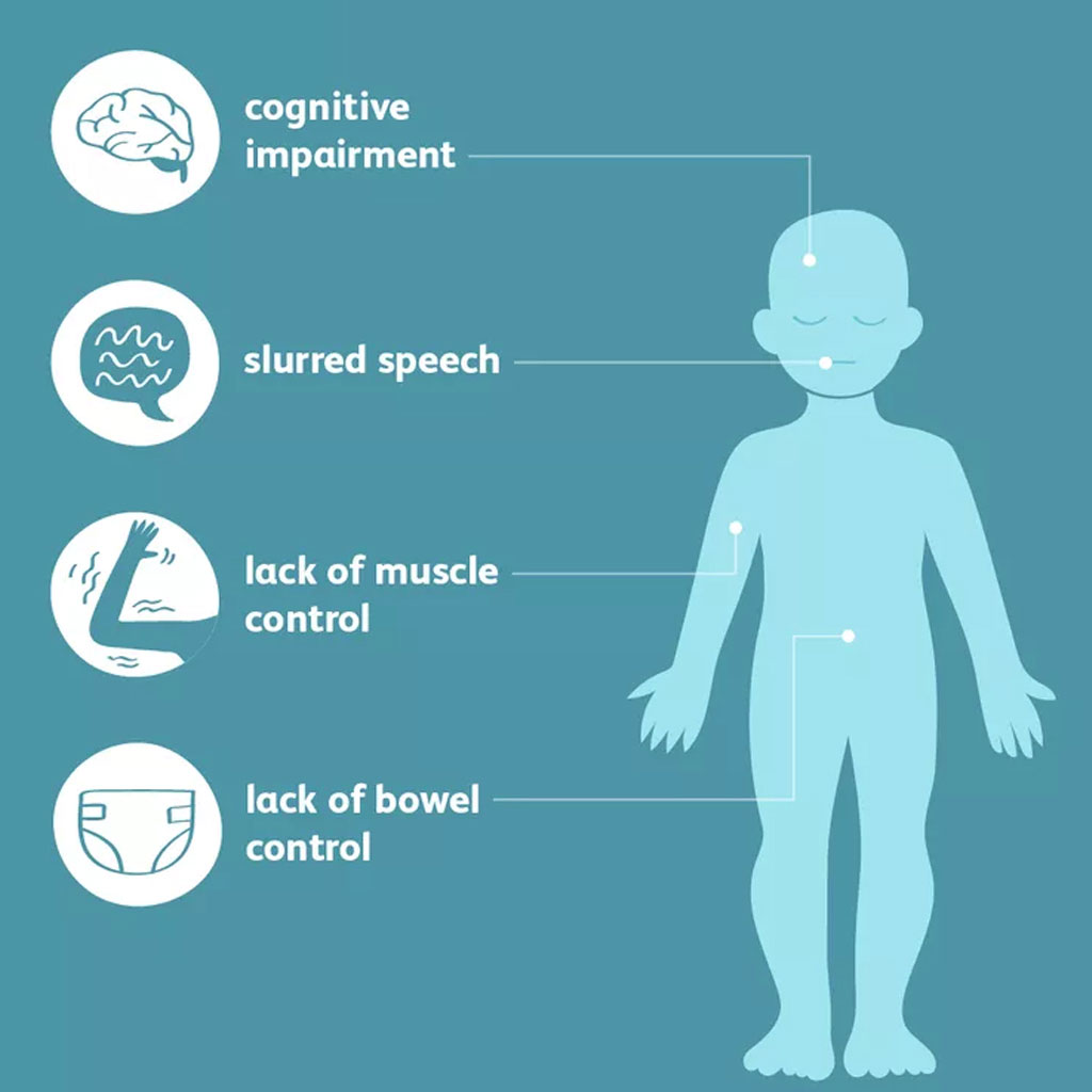
  • Head Injuries: Concussions and more severe traumatic brain injuries are common in children.
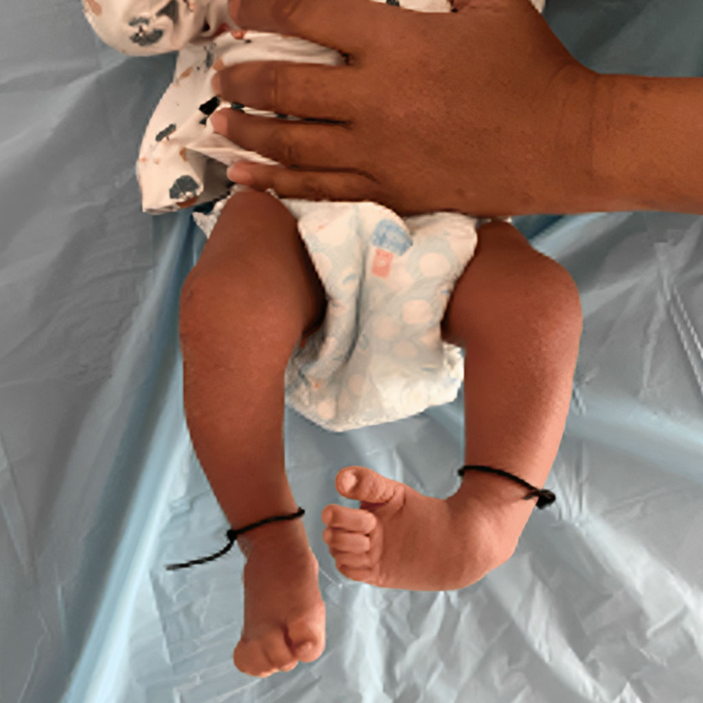
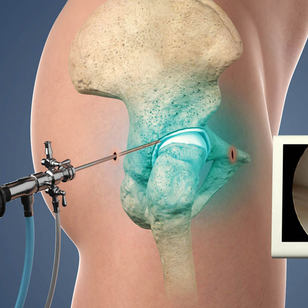
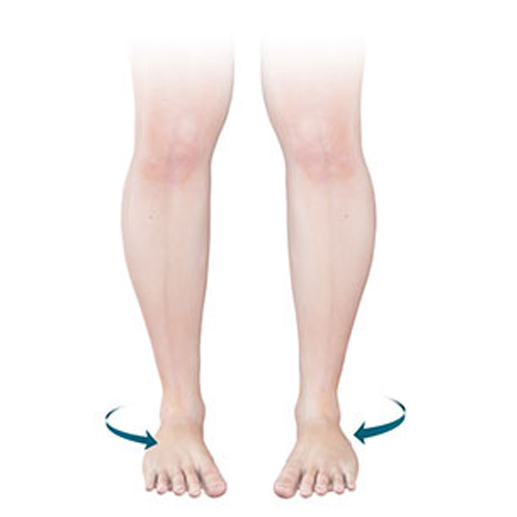
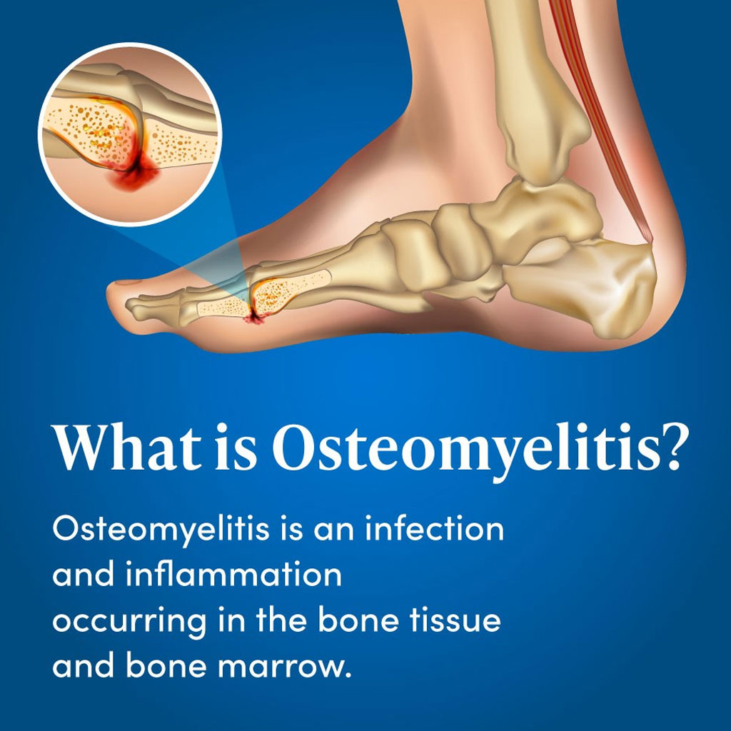
  • Orthopedic Injuries: Fractures, particularly of long bones like the femur, are frequent.
  • Abdominal Trauma: Injuries to internal organs like the spleen or liver can occur in severe cases.
  • Thoracic Trauma: Chest injuries, though less common, can involve the lungs, heart, or ribs.

Postoperative Care and Rehabilitation:

After surgery, rehabilitation often plays a crucial role in helping the child regain full function. Pediatric trauma teams focus on physical therapy, pain management, and emotional support for the child and family.

CTEV

CTEV

Congenital Talipes Equinovarus (CTEV), commonly known as clubfoot, is a congenital deformity of the foot where one or both feet are twisted inward and downward. This condition is present at birth and can vary in severity. Without treatment, clubfoot can cause walking difficulties and permanent disability, but early diagnosis and intervention typically lead to successful correction.

Key Characteristics of CTEV:

  • Foot Deformity: The foot appears turned inward and downward. The affected foot or feet may also be smaller than normal, with underdeveloped calf muscles.

Rigid and Flexible Types

  • Etiology: The exact cause of CTEV is not always clear, but it is believed to involve both genetic and environmental factors. There may be a family history of clubfoot, and certain syndromes or conditions, such as spina bifida, can increase the likelihood of CTEV. It is more common in males than females.

Treatment:

The goal of treatment for CTEV is to correct the position of the foot so the child can walk normally. Early treatment is essential and is typically started shortly after birth. The two most common treatment methods include:

Ponseti Method:

  • Gentle manipulation and casting: The foot is gradually manipulated into the correct position, and a cast is applied to maintain the new position. This process is repeated weekly for several weeks.
  • Tenotomy: In some cases, a minor surgical procedure called a tenotomy (cutting of the Achilles tendon) is performed to release tension in the tendon.
  • Bracing: After the foot is corrected, the child will need to wear a brace (usually at night) to maintain the corrected position and prevent relapse.

French Method (Physical Therapy):

  • Involves daily stretching, manipulation, and taping of the foot, combined with physical therapy. This method requires a more hands-on, long-term approach and active parental involvement.

Surgical Treatment:

  • In cases where conservative methods fail, surgery may be required to correct the foot's structure. Surgery is more commonly used for severe or neglected cases of CTEV.
Cerebral Palsy

Cerebral Palsy

Cerebral Palsy (CP) is a group of neurological disorders that affect a person's ability to move, maintain posture, and balance. It is caused by abnormal brain development or damage to the developing brain, typically before, during, or shortly after birth. CP is the most common motor disability in childhood, and its effects can range from mild to severe.

Causes:

Cerebral palsy results from damage to the areas of the brain that control movement, coordination, and muscle tone. The brain injury is non-progressive, meaning it does not worsen over time, but the symptoms may change as the child grows. Common causes include:

  • Prenatal causes: Lack of oxygen, infections (such as rubella), maternal health issues, or genetic abnormalities.
  • Perinatal causes: Complications during labor and delivery, such as asphyxia (lack of oxygen) or trauma.
  • Postnatal causes: Brain infections (e.g., meningitis, encephalitis), head injury, or stroke in early childhood.

Types of Cerebral Palsy:

Cerebral palsy is classified into different types based on the movement disorder observed:

  • Spastic Cerebral Palsy
  • Dyskinetic (Athetoid) Cerebral Palsy
  • Ataxic Cerebral Palsy
  • Mixed Cerebral Palsy

Symptoms:

The symptoms of CP vary widely depending on the severity and type but may include:

  • Delayed motor milestones: Delayed rolling over, sitting, or walking.
  • Abnormal posture: Children may have unusual or awkward postures due to abnormal muscle tone.
  • Movement difficulties: Stiffness, involuntary movements, or lack of coordination.
  • Balance issues: Difficulty standing or walking independently.
  • Speech and swallowing problems: Difficulty controlling the muscles involved in speaking, eating, and swallowing.

Associated Conditions:

In addition to motor disabilities, many children with cerebral palsy have other associated conditions, including:

  • Seizures (epilepsy).
  • Intellectual disabilities or learning difficulties.
  • Vision or hearing impairments.
  • Speech and language disorders.
  • Orthopedic issues: Scoliosis, joint contractures, or hip dislocation.

Diagnosis:

CP is typically diagnosed based on developmental history, physical examination, and neuroimaging tests such as:

  • MRI or CT scans: These help identify areas of brain damage.
  • Developmental assessments: Tracking the child's motor skills, muscle tone, reflexes, and posture over time.

Treatment:

There is no cure for CP, but early intervention and management can improve quality of life and functional abilities. Treatment is individualized and may involve:

  • Physical Therapy (PT)
  • Occupational Therapy (OT)
  • Speech and Language Therapy
  • Medications
  • Surgery
  • Assistive Technology
Hip Preservation Surgery

Hip Preservation Surgery

Hip preservation surgery refers to a set of surgical procedures aimed at delaying or preventing the need for total hip replacement by preserving the natural hip joint. These surgeries are typically performed on younger patients who have hip conditions that may lead to arthritis or other degenerative issues if left untreated. The goal is to address hip abnormalities, improve function, reduce pain, and prevent further damage to the joint.

Common Conditions Requiring Hip Preservation Surgery:

  • Femoroacetabular Impingement (FAI)
  • Developmental Dysplasia of the Hip (DDH)
  • Hip Labral Tears
  • Hip Dysplasia
  • Perthes Disease
  • Slipped Capital Femoral Epiphysis (SCFE)

Types of Hip Preservation Surgery:

  • Arthroscopy
  • Periacetabular Osteotomy (PAO)
  • Surgical Dislocation and Debridement
  • Proximal Femoral Osteotomy
  • Core Decompression
  • Hip Arthrotomy

Indications for Hip Preservation Surgery:

The symptoms of CP vary widely depending on the severity and type but may include:

  • Chronic hip pain that doesn’t respond to conservative treatments like physical therapy or medications.
  • Limited range of motion or stiffness in the hip.
  • Early signs of osteoarthritis in young or middle-aged adults.
  • Structural abnormalities in the hip joint that are likely to worsen without intervention.

Benefits of Hip Preservation Surgery:

  • Delaying hip replacement: Hip preservation surgeries aim to delay or avoid the need for total hip replacement, particularly in younger patients.
  • Improved function and mobility: By correcting structural issues, patients often experience improved range of motion and less pain.
  • Reduced risk of arthritis: By addressing underlying conditions like FAI or dysplasia, the surgery can help prevent or slow the progression of arthritis.
  • Minimally invasive options: Many procedures, like hip arthroscopy, are minimally invasive, allowing for quicker recovery times compared to open surgeries.
Limb Deformities

Limb Deformities

Limb deformities refer to abnormalities in the shape, structure, or alignment of the bones or muscles in the arms or legs. These deformities can be congenital (present at birth) or acquired (developing after birth due to injury, disease, or other factors). Limb deformities can range from mild, causing minimal functional impairment, to severe, leading to significant mobility issues and physical challenges.

Types of Limb Deformities:

  • Congenital Limb Deformities
  • Acquired Limb Deformities
  • Angular and Rotational Deformities

Causes of Limb Deformities:

  • Genetic Factors
  • Environmental Factors
  • Injury
  • Diseases and Infections

Diagnosis:

  • Physical Examination: A thorough clinical examination to assess the severity and type of deformity.
  • X-rays and Imaging: Used to evaluate bone structure, alignment, and the extent of deformity.
  • CT or MRI: May be used for complex cases, especially when soft tissue or joint involvement is suspected.
  • Gait Analysis: In some cases, a detailed analysis of walking patterns is performed to assess functional impairment and guide treatment.

Treatment of Limb Deformities:

Treatment varies based on the severity, type, and cause of the deformity. The primary goals are to restore normal function, correct appearance, and prevent further complications.

Non-Surgical Treatments:

  • Bracing: Used for milder deformities like bowlegs or knock knees to guide the limbs into proper alignment as the child grows.
  • Physical Therapy: Focuses on improving muscle strength, flexibility, and movement to help manage mild deformities.
  • Corrective Casting: In infants, deformities like clubfoot can be treated using casting techniques, such as the Ponseti method.
  • Orthotics: Special shoes, insoles, or braces may be prescribed to improve function and support alignment.

Surgical Treatments:

  • Osteotomy: A surgical procedure where the bone is cut and realigned to correct angular deformities like bowlegs or knock knees.
  • Limb Lengthening: For significant limb length discrepancies, an external fixator may be used to slowly stretch the bone and stimulate new growth.
  • Epiphysiodesis: In cases of growth plate injuries or discrepancies in limb length, this procedure involves intentionally stopping the growth of a longer limb to allow the shorter limb to catch up.
  • Excision or Amputation: In cases where a deformed or non-functional limb is present, part or all of the limb may be surgically removed.
  • Reconstructive Surgery: This may involve tendon transfers, bone grafts, or joint realignment to restore function.

Minimally Invasive Techniques:

  • Arthroscopy: For some joint-related deformities, arthroscopy can be used to correct structural abnormalities in a less invasive way.
Foot Deformities

Foot Deformities

Foot deformities are abnormalities in the structure, shape, or alignment of the bones, muscles, or ligaments of the foot. These deformities can be congenital (present at birth) or acquired due to injury, disease, or other factors. Foot deformities can cause pain, mobility issues, and affect the overall function of the foot, leading to difficulties with walking or standing.

Types of Foot Deformities:

  • Congenital Foot Deformities
  • Acquired Foot Deformities
  • Toe Deformities

Causes of Foot Deformities:

  • Genetic Factors: Many congenital foot deformities, such as clubfoot or polydactyly, are linked to genetic factors or hereditary conditions.
  • Injury or Trauma: Improper healing of fractures or ligament injuries can lead to acquired deformities such as flatfoot or hammertoe.
  • Poor Footwear: Wearing high heels, narrow shoes, or shoes that don’t fit properly can contribute to bunions, hammertoes, and other deformities.
  • Neurological Disorders: Conditions such as cerebral palsy, Charcot-Marie-Tooth disease, or stroke can affect muscle control, leading to deformities like pes cavus or claw toe.
  • Arthritis: Inflammatory conditions such as rheumatoid arthritis can cause joint damage and deformities, including bunions or hammertoes.
  • Diabetes: Diabetes can lead to nerve damage (peripheral neuropathy), which in turn can contribute to conditions like Charcot foot.

Diagnosis:

  • Physical Examination: The doctor will observe the foot structure, range of motion, and gait.
  • X-rays: Used to visualize bone alignment and assess the severity of deformities.
  • MRI or CT scans: May be used in complex cases, especially when soft tissue involvement is suspected.
  • Gait Analysis: Sometimes performed to evaluate walking patterns and the impact of the deformity on movement.

Treatment of Foot Deformities:

Conservative (Non-Surgical) Treatment:

  • Orthotics: Custom-made shoe inserts can help support the foot, relieve pressure, and improve alignment in cases of flatfoot, bunions, or high arches.
  • Bracing: In conditions like clubfoot, corrective braces can help maintain foot positioning after initial treatment (e.g., the Ponseti method).
  • Physical Therapy: Stretching, strengthening, and range-of-motion exercises can help improve function and prevent worsening of deformities like hammertoes or flatfoot.
  • Proper Footwear: Wearing supportive, well-fitted shoes can help manage conditions like bunions and prevent further deformities.
  • Medications: Anti-inflammatory drugs can reduce pain and swelling in conditions like arthritis.

Surgical Treatments:

  • Bunionectomy: Removal of the bony bump on the side of the foot, often combined with realigning the big toe.
  • Tendon Transfer: In cases of claw toe or high-arched foot, tendon transfers can help restore muscle balance.
  • Osteotomy: Cutting and repositioning bones to correct alignment in deformities like severe bunions or flatfoot.
  • Arthrodesis (Joint Fusion): Fusing certain joints in the foot to correct deformities and reduce pain in cases of severe arthritis or unstable foot structures.
  • Excision of Extra Digits: Surgical removal of extra toes (polydactyly) to improve function and appearance.
  • Clubfoot Repair: In some cases, surgery is needed to release tight tendons and reposition the bones.
Osteomyelitis

Osteomyelitis

Osteomyelitis is a serious bone infection caused by bacteria or, less commonly, fungi. It can affect any bone in the body, but it most frequently occurs in the long bones of the arms and legs, the spine, and the pelvis. Osteomyelitis can occur at any age and may develop suddenly (acute) or slowly over time (chronic).

Causes of Osteomyelitis:

  • Hematogenous spread: Bacteria or fungi from an infection elsewhere in the body (such as in the skin, lungs, or urinary tract) enter the bloodstream and spread to the bone.
  • Direct inoculation: The infection enters the bone directly from an open wound or surgery, often due to fractures, orthopedic surgery, or trauma.
  • Contiguous spread: The infection spreads from nearby infected tissue or an open wound (e.g., diabetic foot ulcer or pressure sores).

Common Pathogens:

  • Staphylococcus aureus (especially methicillin-resistant Staphylococcus aureus, MRSA) is the most common cause.
  • Streptococcus species and Pseudomonas aeruginosa are also frequently implicated.
  • In immunocompromised individuals, fungi and other atypical organisms can be responsible.

Symptoms of Osteomyelitis:

  • Bone pain: The hallmark symptom, which may be severe and worsen with movement.
  • Swelling, redness, and warmth over the affected area.
  • Fever and chills: Especially in acute osteomyelitis.
  • Fatigue and malaise: General feeling of being unwell.
  • Limited range of motion in the affected limb or joint.
  • Drainage from a sinus tract: In chronic osteomyelitis, a fistula may form, allowing pus to drain from the bone to the skin.

Diagnosis:

  • Physical Examination: The doctor will check for tenderness, swelling, and redness over the affected area.

Blood Tests:

  • Complete blood count (CBC): Elevated white blood cell count in response to infection.
  • Erythrocyte sedimentation rate (ESR) and C-reactive protein (CRP): Inflammation markers that are typically elevated in osteomyelitis.
  • Blood cultures: Help identify the bacteria or fungi causing the infection if hematogenous spread is suspected.

Imaging:

  • X-rays: Early in the disease, X-rays may be normal, but over time, they can show bone destruction and new bone formation.
  • MRI: More sensitive than X-rays, can detect early changes in bone and surrounding tissues.
  • CT scan: Useful for evaluating bone destruction in chronic osteomyelitis.
  • Bone scan: A nuclear imaging test that detects areas of increased bone activity, often used when MRI is unavailable.

Bone Biopsy:

  • A bone biopsy is the gold standard for diagnosing osteomyelitis. It involves taking a sample of bone tissue to identify the exact organism causing the infection and determine its antibiotic sensitivity.

Treatment of Osteomyelitis:

Antibiotic Therapy:

  • The cornerstone of treatment, especially for bacterial osteomyelitis.
  • Antibiotics are typically administered intravenously for 4-6 weeks, followed by oral antibiotics.
  • The choice of antibiotics depends on the organism identified by blood cultures or biopsy (e.g., MRSA might require vancomycin).
  • In some cases, long-term oral antibiotics are necessary to prevent recurrence.

Surgical Treatment:

  • Debridement: Removal of infected or dead bone tissue (sequestrum) and soft tissue.
  • Drainage of abscesses: If pus has accumulated, it may need to be drained surgically.
  • Bone grafting: After infected tissue is removed, bone grafts or substitutes may be used to fill defects and promote healing.
  • Stabilization of bones: In some cases, surgical hardware (e.g., plates, screws) may be required to support the affected bone, especially if there is structural weakness.

Chronic Osteomyelitis Management:

  • Requires long-term antibiotics and may involve multiple surgeries to manage recurrences and remove dead tissue.
  • Sinus tracts may need to be surgically removed to prevent re-infection.
  • In extreme cases, amputation of the affected limb may be necessary if infection cannot be controlled.

Hyperbaric Oxygen Therapy (HBOT):

  • Used as an adjunct treatment in certain cases of chronic osteomyelitis or when healing is impaired (e.g., in diabetic foot osteomyelitis).
  • Involves breathing pure oxygen in a pressurized environment, which enhances the ability of blood to deliver oxygen to the infected tissues and promotes healing.
Benign Tumors

Benign Tumors

Benign tumors are non-cancerous growths that arise when cells divide and grow excessively but do not invade surrounding tissues or spread to other parts of the body (metastasis). While benign tumors are generally less harmful than malignant (cancerous) tumors, they can still cause issues depending on their size, location, and impact on nearby structures.

Characteristics of Benign Tumors:

  • Non-invasive: They grow locally without spreading to other tissues.
  • Slow-growing: Benign tumors tend to grow slowly compared to malignant ones.
  • Encapsulated: They are often enclosed in a fibrous capsule, making them easier to remove surgically.
  • No metastasis: Unlike cancerous tumors, benign tumors do not spread to distant parts of the body.
  • Well-differentiated: Cells within benign tumors resemble normal cells and maintain their function to a degree.

Common Types of Benign Tumors:

  • Lipomas
  • Fibromas
  • Hemangiomas
  • Adenomas
  • Osteochondromas
  • Nevi (Moles)
  • Meningiomas
  • Schwannomas
  • Chondromas
  • Dermoid Cysts

Causes and Risk Factors:

  • Genetic mutations: Some benign tumors result from mutations in specific genes that control cell growth and division.
  • Hormonal influences: Hormonal changes or imbalances, such as estrogen levels, can promote the growth of certain benign tumors (e.g., uterine fibroids).
  • Inflammation or injury: In some cases, chronic inflammation or injury to tissue may contribute to benign tumor formation.

Symptoms of Benign Tumors:

  • Local swelling or lump: Most benign tumors are discovered as lumps or swellings.
  • Pain or discomfort: Some tumors may press on nearby nerves, organs, or tissues, causing pain or discomfort.
  • Functional impairment: Depending on the location, benign tumors can interfere with normal bodily functions (e.g., brain tumors causing neurological symptoms).
  • Cosmetic concerns: Tumors on the skin or visible areas can cause cosmetic issues.

Diagnosis:

  • Physical Examination: The doctor will check for lumps or masses, assess their characteristics, and evaluate any symptoms.
  • Biopsy: A tissue sample may be taken to determine whether the tumor is benign or malignant. The biopsy can be done through a needle or during surgery.

Treatment of Benign Tumors:

  • Observation: Many benign tumors do not require treatment unless they cause symptoms or there’s a risk of malignancy.
  • Surgical Removal: Surgery is often performed if the tumor is causing discomfort, impairing function, or for cosmetic reasons. In some cases, tumors are removed to prevent the possibility of malignancy (e.g., colon adenomas).
  • Medications: Hormonal therapies or medications can sometimes shrink tumors (e.g., in the case of uterine fibroids).
  • Radiation Therapy: Rarely used for benign tumors, but may be employed in cases like meningiomas where surgery is not feasible.
Deformity Correction

Deformity Correction

Deformity correction is a surgical or non-surgical approach used to correct abnormalities in the alignment, shape, or length of bones or joints. These deformities can result from congenital conditions, trauma, infections, tumors, or developmental abnormalities and may lead to functional impairment, pain, or aesthetic concerns.

Types of Deformities:

  • Congenital Deformities: Present at birth, such as clubfoot (congenital talipes equinovarus), congenital hip dysplasia, or scoliosis.
  • Acquired Deformities: Develop later in life due to injury, infection, arthritis, or diseases such as rickets or osteomyelitis.
  • Post-Traumatic Deformities: Result from improperly healed fractures (malunion), which may cause misalignment of bones or joints.
  • Degenerative Deformities: Caused by conditions like osteoarthritis or rheumatoid arthritis, leading to joint destruction and deformity over time.

Goals of Deformity Correction:

  • Restoration of Function: Correcting deformities to improve the ability to walk, use hands, or perform daily activities without pain or restriction.
  • Pain Relief: Realigning bones or joints to reduce abnormal stresses that lead to pain.
  • Prevention of Joint Degeneration: Preventing long-term complications like arthritis by correcting malalignment early.
  • Improving Aesthetics: Restoring normal appearance and symmetry, particularly important in facial and limb deformities.
  • Enhanced Quality of Life: Increased mobility, independence, and confidence following correction.

Devices Used in Deformity Correction:

  • External Fixators (e.g., Ilizarov or Taylor Spatial Frame)
  • Internal Fixation (Plates, Screws, Nails)
  • Growth Modulation Devices

Post-Surgical Care and Rehabilitation:

  • Physical Therapy: Vital for restoring strength, flexibility, and function after surgery. Patients often undergo rehabilitation to regain movement and learn how to use the corrected limb or joint.
  • Pain Management: Includes medications and other modalities like ice, compression, and elevation.
  • Weight-bearing Restrictions: In cases of limb surgeries, patients may need to avoid bearing weight on the affected limb for weeks or months while healing.
  • Follow-up Imaging: X-rays or other imaging modalities are used to monitor bone healing and ensure proper alignment is maintained.

Complications of Deformity Correction Surgery:

  • Infection: Particularly with external fixators, there is a risk of infection at the pin sites.
  • Non-union or Malunion: The bone may fail to heal properly, requiring further intervention.
  • Nerve or Blood Vessel Injury: Due to the proximity of these structures during surgery.
  • Joint Stiffness: Post-surgical stiffness may require additional therapy or procedures to improve mobility.

Indications for Surgery:

  • Severe Functional Impairment: When a deformity causes significant limitations in daily activities.
  • Progressive Deformity: Conditions like scoliosis or limb length discrepancy that worsen over time.
  • Persistent Pain: When conservative treatments like bracing or therapy fail to relieve symptoms.
  • Cosmetic Concerns: Some patients seek correction for aesthetic reasons, especially with visible deformities.
Congenital Anomalies

Congenital Anomalies

Congenital anomalies, also known as birth defects, are structural or functional abnormalities present at birth that can affect various parts of the body. These anomalies can arise from genetic, environmental, or unknown causes and may range from minor to severe. They can affect the organs, limbs, or overall body structure and can be isolated or part of a syndrome.

Causes and Risk Factors

  • Genetic Factors
  • Environmental Factors
  • Unknown Factors

Diagnosis

Prenatal Screening:

  • Ultrasound: Can detect structural abnormalities in the fetus, such as heart defects or neural tube defects.
  • Maternal Serum Screening: Blood tests to assess risk factors for certain conditions (e.g., Down syndrome).
  • Amniocentesis: Sampling of amniotic fluid to test for genetic conditions.
  • Chorionic Villus Sampling (CVS): Sampling of placental tissue for genetic testing.

Postnatal Diagnosis:

  • Physical Examination: Identifying visible physical abnormalities.
  • Imaging Studies: X-rays, CT scans, MRI for internal anomalies.
  • Genetic Testing: Blood tests or tissue samples to identify specific genetic disorders.
  • Metabolic Screening: Newborn screening tests to detect metabolic disorders.

Management and Treatment

Medical Management:

  • Medications: For conditions like congenital hypothyroidism or cystic fibrosis.
  • Hormone Therapy: For conditions like congenital adrenal hyperplasia.

Surgical Intervention:

  • Repair or Correction: Many structural anomalies require surgical correction, such as cleft lip/palate repair, heart defect surgery, or limb deformity correction.

Therapeutic Interventions:

  • Physical Therapy: For improving motor skills and functional abilities in conditions like cerebral palsy.
  • Occupational Therapy: To assist with daily living activities and adaptive skills.
  • Speech Therapy: For speech and language development issues, especially in conditions like cleft palate.

Specialist Care:

  • Multidisciplinary Teams: Involvement of various specialists including pediatricians, surgeons, geneticists, and therapists to provide comprehensive care.

Supportive Care:

  • Educational Support: For developmental or learning disabilities.
  • Counseling: Psychological support for the child and family to cope with chronic conditions or long-term disabilities.
Pre Natal Counselling

Pre Natal Counselling

Prenatal counseling is an essential part of prenatal care that provides prospective parents with information, guidance, and support to prepare for the health and well-being of both the mother and the baby. It involves discussing various aspects of pregnancy, childbirth, and parenting, as well as assessing and managing any potential risks or concerns.

When to Start Prenatal Counseling

  • Early in Pregnancy: Ideally, prenatal counseling should begin as soon as pregnancy is confirmed, allowing ample time to address all relevant topics and make informed decisions.
  • Ongoing: Counseling may continue throughout the pregnancy as new issues or questions arise.

Benefits of Prenatal Counseling

  • Informed Decisions: Helps parents make informed choices about prenatal care, testing, and delivery options.
  • Early Detection: Identifies potential health issues early, allowing for timely intervention.
  • Emotional Support: Provides reassurance and support, helping to alleviate stress and anxiety.
  • Better Outcomes: Promotes healthier behaviors and practices, leading to better outcomes for both mother and baby.

Common Challenges and Solutions

  • Understanding Complex Information: Medical jargon and complex information can be overwhelming. Counselors should use clear, understandable language and provide written materials.
  • Emotional Reactions: Dealing with the possibility of health issues or genetic conditions can be emotionally challenging. Providing compassionate support and referrals to mental health professionals can help.
  • Cultural Sensitivity: Respecting cultural beliefs and practices related to pregnancy and childbirth is crucial. Tailoring counseling to meet cultural preferences can improve acceptance and adherence.